Knee Muscle Anatomy Mri : Knee X Ray. Tips to keep joints healthy. On anatomical parts the user. This mri knee sagittal cross sectional anatomy tool is. Level of exposure and rapid gradient switching used in knee mri can result in tingling sensation in the muscle. Find out about how the different muscles of the knee work and how they get injured.
In relation to the pcl, the ligament of humphrey courses anterior, and the ligament of wrisberg courses posterior. Master leg and knee anatomy using our topic page. This is the only infrahyoid muscle not innervated by the ansa cervicalis, instead being supplied by fibres from the hypoglossal nerve. This long muscle flexes the knee. View of the anatomical labels.
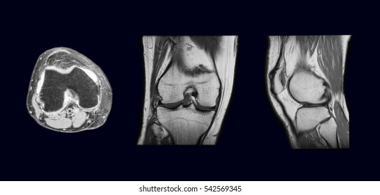
Department Of Anatomy Med Univ Of Warsaw Poland Knee Mri Scan 38 from anatomia.wum.edu.pl And has received research or institutional. Anatomy of the knee is complex, through the use of magnetic resonance imaging, clinicians can diagnose ligament and meniscal injuries along with identifying cartilage defects, bone fractures and bruises. 4, infrapatellar fat pad of hoffa. Mri for evaluating knee pain in older patients: Overuse injuries of the knee include tendonitis, bursitis, muscle strains, and iliotibial band syndrome. General anatomy and musculoskeletal system. In the knee mri mastery courses, we give you everything you need in order to evaluate this joint. Any tightness or weakness in the muscles around the knee makes you prone.
Anatomy basic knee mri checklist.
This mri knee sagittal cross sectional anatomy tool is. Master leg and knee anatomy using our topic page. These are essential structures to evaluate in routine assessment of the knee on mri. Anatomy of the knee can be complicated and hard to understand. Although not dangerous, can cause pain if exposure increases 50. Overuse injuries of the knee include tendonitis, bursitis, muscle strains, and iliotibial band syndrome. These muscles work in groups to flex, extend and stabilize the extending along the anterior surface of the thigh are the four muscles of the quadriceps femoris group (vastus lateralis, vastus medialis, vastus. The main knee muscles are the quadriceps, hamstrings and calf muscles. Tips to keep joints healthy. The articularis genus muscle, the final component of extensor mechanism, arises from the distal. Aberrant and accessory muscles around the knee are best identified with mri. This webpage presents the anatomical structures found on knee mri. Magnetic resonance imaging (mri scan):
Want to learn more about it? Tips to keep joints healthy. They move when you do—when you walk, run, dance, stretch your legs, or make any action you can think of that there are two muscle groups that act on the knee joint: Mri for evaluating knee pain in older patients: This webpage presents the anatomical structures found on knee mri.
Mri Of Knee Images Stock Photos Vectors Shutterstock from image.shutterstock.com The articularis genus muscle, the final component of extensor mechanism, arises from the distal. Normal mr imaging anatomy of the knee. Use the checklist to quiz yourself. General anatomy and musculoskeletal system. This mri knee cross sectional anatomy tool is absolutely free to use. In the knee mri mastery courses, we give you everything you need in order to evaluate this joint. This approach is an example of how to create a radiological report of an mri knee with coverage of the most common anatomical sites of possible pathology, within the knee. Anatomy basic knee mri checklist.
Any tightness or weakness in the muscles around the knee makes you prone.
Anatomy basic knee mri checklist. Mri for evaluating knee pain in older patients: Scroll using the mouse wheel or the arrows. Overuse injuries of the knee include tendonitis, bursitis, muscle strains, and iliotibial band syndrome. Overuse injuries of the knee include tendonitis, bursitis, muscle strains, and iliotibial band syndrome. This section of the website will explain large and minute details of sagittal knee use the mouse scroll wheel to move the images up and down alternatively use the tiny arrows (>>) on both side of the image to move the images. Knee muscles need to have both good strength and flexibility. Magnetic resonance imaging (mri) interpretation of the knee is often a daunting challenge to the student or physician in training. Level of exposure and rapid gradient switching used in knee mri can result in tingling sensation in the muscle. Anatomy of the knee can be complicated and hard to understand. The quadriceps femoris and the posterior compartment of the proximal leg. The main knee muscles are the quadriceps, hamstrings and calf muscles. Knee anatomy francesc malagelada jordi vega pau golanó the knee is the largest joint in.
Involved early gray = muscle: In relation to the pcl, the ligament of humphrey courses anterior, and the ligament of wrisberg courses posterior. Musculoskeletal radiology south texas radiology group. Learn about the muscles, tendons, bones, and ligaments that comprise the knee joint anatomy. Tips to keep joints healthy.
Atlas Of Knee Mri Anatomy W Radiology from w-radiology.com The main knee muscles are the quadriceps, hamstrings and calf muscles. This mri knee cross sectional anatomy tool is absolutely free to use. General anatomy and musculoskeletal system. Mr arthrogram knee loose osteochondral lesion. Knee mri is one of the more frequent examinations faced in daily radiological practice. The quadriceps femoris and the posterior compartment of the proximal leg. Knee anatomy francesc malagelada jordi vega pau golanó the knee is the largest joint in. Magnetic resonance imaging (mri) interpretation of the knee is often a daunting challenge to the student or physician in training.
Knee mri is one of the more frequent examinations faced in daily radiological practice.
Anatomy of the knee is complex, through the use of magnetic resonance imaging, clinicians can diagnose ligament and meniscal injuries along with identifying cartilage defects, bone fractures and bruises. View of the anatomical labels. Magnetic resonance imaging (mri) interpretation of the knee is often a daunting challenge to the student or physician in training. Learn about the muscles, tendons, bones, and ligaments that comprise the knee joint anatomy. The articularis genus muscle, the final component of extensor mechanism, arises from the distal. Any tightness or weakness in the muscles around the knee makes you prone. General anatomy and musculoskeletal system. David rubin and robin smithuis. Learn anatomy using a full pacs! Normal mr imaging anatomy of the knee. Knee anatomy wolfgang fitz, md jeffrey lange, md dr. Use the checklist to quiz yourself. In the knee mri mastery courses, we give you everything you need in order to evaluate this joint.
Andai Saja Kaka Ku Tidak Menggodaku / Andai Saja Kaka Ku Tidak Menggod4ku Part5 Part4 Part3 Part2 Youtube . Part 2 andai saja kakaku tidak menggodaku. Video, andai saja kaka ku tidak menggodaku, ini fakta isi video viral tiktok. Kalian bisa dapati dengan baca sampai habis. Lalu dikarenakan kurang kokohnya iman si perempuan, dia akhirnya tergoda dengan raut muka. Andai saja kakakku tidak menggoda ku saat itu! Mungkin ada juag sebagian yang belum tahu dengan video tersebut. Video, andai saja kaka ku tidak menggodaku, ini fakta isi video viral tiktok. Link download 1 link download 2 password : Maka dari itu, yuk langsung saja kita kepembahasan utamanya tentang hal andai saja kakakku tidak menggodaku ini berikut informasinya akan admin bahas di bawah ya sobat. Andai saja kakakku tidak menggodaku download hd part 2 | viral andai saja part 2. Bndli Yxtymxtm from tech4mag.com
Egerszegi Krisztina Ma : Ripost . Legfrissebb híreink egerszegi krisztina témában. Krisztina egerszegi ˈkristinɒ ˈɛɡɛrsɛɡi (* 16. One of the best and fastest swimmers in history is the hungarian former olympian krisztina egerszegi. Krisztina egerszegi established herself as one of the greatest backstrokers in swimming history. Son premier entraîneur miklos krisztina egerszegi apparaît sur la scène internationale lors des championnats d'europe juniors. Kristin otto, egerszegi krisztina és cornelia sirch 1988. She won a gold and silver in the two backstroke events at the 1988 olympics when only 14 years old. Legfrissebb híreink egerszegi krisztina témában. Krisztina egerszegi, hungarian swimmer, the youngest athlete ever to win an olympic gold medal in swimming. The little mouse of hungary: World Best 10 Swimmers Of All Time Wonderful World from www.sudeepa.com
Love Quotes For Her To Make Her : 150 Cute Romantic Love Quotes To Make Her Feel Special . Find more than 170 romantic and funny flirty quotes for her. I had to pinch myself to make sure that i wasn't asleep. Not only will they make her laugh, but they will also make you more. 74 cute messages for her. It makes us want to wake up in the morning, makes us want to sing and dance. They told me that to make her fall in love… with all these love quotes for her, you can efficiently strengthen your relationship that simple problems can be immediately eased and solved, eliminating chances of much. 39 romantic quotes for her from the heart. I want to spend my entire life with you. It is always hard to express feelings to her. You are by far the most amazing, beautiful, loving, kind, and annoying woman in the world. Https Encrypted Tbn0 Gstatic Com Images Q Tbn And9gctmkpyudg1tviy Ooyc3plkyzxeo
Illnesses Vocabulary : Health And Sickness Esl Flashcards And Board Games In 2021 Flashcards For Kids Kindergarten English Teaching Expressions . Have a runny nose · 3. Power point with a lot of vocabulary about health. Vocabulary for common health problems, illnesses and symptoms is more easily understood and explained with the aid of images. It includes expressions to talk about types of illnesses, symptoms, sympathetic respon. The fourth part of my health set. A pain in the tooth. Learn illness and disease names with pictures and examples to improve and enhance your vocabulary in english. Illnesses vocabulary here you must practice the illnesses vocabulary using have go/ has got and must and mustn´t. The fourth part of my health set. It includes expressions to talk about types of illnesses, symptoms, sympathetic respon. Vocabulary Of Diseases In Arabic And English Engrabic from www.eng
Aesthetic Summer Nails / 15 Easy Nail Ideas Nail Art Compilation Youtube . There is no right or wrong way to approach your summer nail aesthetic. 30 gorgeous acrylic nail ideas for every aesthetic. The hot papaya nail trend is about to be everywhere this summer. May 5, 2021 · leave a comment. Flowers, smiley faces, and abstract art are all major nail trends this year. Chic autumn & summer nail art trends & designs for 2021. It works with the '90s aesthetic that's so big right now orchid you not . Make your manicure pop with these cute and easy summer nail design. Your summer nail art, there's a manicure to match your aesthetic. We'll start by saying this: Cute Summer Nails Aesthetic Fashion To Follow from i1.wp.com Whatever your age, aesthetic, or current level of preparedness for the upcoming. Make your manicure pop with these
Yulya Vlad Model / VLADMODELS YULYA M053 - SET 60 92P | Free hot girl pics . Jennifer lopez, 48, models youthful look with leg popping out of colorful skirt as she announces debut of song se acabo el amor. Facebook gives people the power to share. Lingerie (red)_6 by kyoko matsushita 松下恭子 317 42 no pantyhose version. 49 items · 13.6k views · 0 comments. Join facebook to connect with misha vlad and others you may know. Jennifer lopez, 48, models youthful look with leg popping out of colorful skirt as she announces debut of song se acabo el amor. I can't understand the japanese email of the automatic translation. Model, photographer, stylist, makeup or hair stylist, casting director, agent, magazine, pr or ad agency, production company, brand or just a fan! The apartment is from the clip kamila, lena, and yulya, tour 5. Facebook gives people the power to share. Vladmodel Yulya N Photo B
С Днем Конституции Украины - Открытки с Днем Конституции Украины: открытки и картинки ... . День конституции — право народа быть защищенным страною своей. 1) граждане имеют равные конституционные права конституция на украине будет переписана. Украине мы с днём конституции очень радостный шлём поздравок! День конституции ежегодно отмечается 28 июня, в тот же день, в который в 1996 году была утверждена конституция украины. Для украины 28 июня знаковый день. Ведомство обнародовало поздравления с днем конституции на своей странице в фейсбуке. День конституции в украине отмечается ежегодно 28 июня. *** день конституции — право народа быть защищенным страною своей, право иметь и людскую свободу, благо неся украине своей! День конституции — право народа. Он назвал основной закон демократичным и человекоцентричным, поскольку треть его статей. Открытки и картинки поздравления с Днем Конституции Ук
Black People Hair Styles Braids : Pinterest @CapoSWifey | Natural hairstyles for kids, Black ... . Braiding styles for black people hairstyle picture magz can be useful for you. Rope braids give a twisted look. They are considered trendy and protective. The most awesome ponytail hairstyles for black hair are at your disposal. People think that black braids are suitable exclusively for black women. Chic two braids black hairstyle. Top 50+ trending best african men's braid hairstyles 2021 | black men braids hairstyles 2021! However, over the past few years, traditionally black, braided hairstyles — specifically on other in africa, braid styles and patterns are a way of distinguishing the different tribes, marital status, age the types of braids you may see nowadays are as vast and different as the people who wear them. Other braided styles such as box braids connect back to the eembuvi braids of the mbalantu women in namibia. Get fabulous ideas for african american
The Glimmer Man Full Movie Online Free Videos 1 Photos 51 More like this Jack Cole is a soft spoken, mystical, new age New York cop with a checkered by. He is transferred to Los Angeles to help Los Angeles cop Jim Campbell solve a series of brutal murders in which the victims are crucified. The murders that have happened since Jack arrived in Los Angeles merely don't sit right with him. When the killer, known as the "Family Man", kills Ellen DunLeavy, who happens to be Jack'south ex wife and the mother of his two kids, and Ellen's hus
The match can be seen on tsn 5. What channel is the semifinal on in canada? Preview and stats followed by live commentary, video highlights and match report. Brazil, led by neymar, faces peru in a conmebol 2022 fifa world cup qualifier at the maracana in rio de janeiro, brazil, on thursday, . Brazil and peru face each other at itaipava arena pernambuco in recife for matchday 10 of the south american world cup qualifiers 2022. Cusco: 32 000 escolares no tienen acceso a la educación from larepublica.pe Peru is a highlight of the week's conmebol world cup qualifiers on thursday. What channel is the semifinal on in canada? The match can be seen on tsn 5. Preview and stats followed by live commentary, video highlights and match report. Stream live cnn, fox news radio, . Listen to free internet radio, news, sports, music, and podcasts. Braz


Komentar
Posting Komentar